Friday, May 3, 2024

Sudden shock with a Nasty looking ECG. What is it?

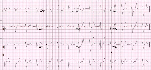
A 60-something woman complained of sudden severe abd pain. She was found by medics agitated, hypotensive, diaphoretic, and in shock.


There were 2 prehospital ECGs:

What do you think?


Smith: Uncertain supraventricular rhythm with PVCs. (See Ken Grauer's analysis below). There is "shart fin" in I and aVL, which is due to a combination of a large R-wave due to left anterior fascicular block plus downsloping ST elevation due to OMI.  There is reciprocal STD in inferior leads.  There is a rather large R-wave in lead V1 and a very large R-wave in V2, suggesting an atypical RBBB.  There is huge ST depression across the precordial leads.  There is STE in aVR.  Thus, there is high lateral OMI with diffuse ST depression.  

When I was shown this ECG, I said it looks like such widespread ischemia that is might be a left main occlusion, or LM ischemia plus circumflex occlusion (high lateral and posterior OMI).  Moreover, left main occlusion often presents near death.  In fact, most do not make it to the hospital alive, which explains why only a tiny percent of OMI are due to full LM occlusion.

Here is the Queen of Hearts interpretation:


There is a second prehospital ECG:

Again, supraventricular rhythm with RBBB and LAFB, shark fin, and STD maximal in V3.  Posterior and high lateral OMI.
But this time the Queen gets it wrong (thinks it is not OMI):


There were runs of VT:


Tha patient arrived in profound shock and had an ED ECG:

Now there is some evolution to include the ST elevation (rather than ST depression) in V4-V6.  This suggests that thrombus has propagated to occlude the LAD in addition to the circumflex.


It can be difficult to see ST elevation and depression when there is RBBB.  
--It helps to find the end of the QRS.  
--This is most easily done in lead V1.  
--Then you can draw a line down through leads V2 and V3 to the lead II rhythm strip across the bottom.  
--Then you can find the same location on the QRS in each 2.5 second interval.  
--Then you can draw the line up to each of leads I, II, III; then aVR, aVL, aVF; then V4, V5,V6.


The Queen of Hearts gets it right here:


The interventionalist stated that he could not do the procedure while the patient has a blood pressure of 45 systolic.

Therefore, the patient was put on arterio-venous ECMO.

Then an angiogram was done.

There was a 100% Left Main Occlusion (OMI).  It was opened and there was thrombus in the circ and LAD.

Unfortunately, the patient ultimately died.

Learning Points:

1. RBBB + LAFB in the setting of ACS is very bad.  Some patients have baseline RBBB with LAFB, but in patients with likely ACS, these are associated with severe infarction with cardiac arrest, cardiogenic shock or impending shock.

2. Patients with ACS and RBBB/LAFB usually have a left main vs. proximal LAD. 

Here are some cases of RBBB with LAFB: 

What is the Diagnosis in this 70-something with Chest Pain?


3. Left Main Non-Occlusive ACS presents with widespread ST Depression and STE in aVR.  Total Left Main Occlusion presents with different ECG findings which are multi-faceted.

See the variety of Left Main Occlusion ECGs here: 

How does Acute Total Left Main Coronary occlusion present on the ECG?


===================================

MY Comment, by KEN GRAUER, MD (5/1/2024):

===================================
Most patients with acute LMain Occlusion do not survive to make it to the hospital. Today's patient did make it to the hospital — but was in cardiogenic shock, and despite valiant attempt at treatment, succumbed soon after.
  • I focus my comment on some additional Learning Points to those highlighted by Dr. Smith.


ECG Findings with Acute LMain Occlusion:
As per Dr. Smith — the ECG findings of total acute LMain occlusion are multifaceted. In Figure-1 — I reproduce major points that I've summarized from Dr. Smith's August 9, 2019 post on the subject. The KEY Take Home Points are as follows:
  • There is no “single” ECG presentation for patients with acute LMain occlusion. Quite literally — You can see almost anything!
  • The reason for this highly variable ECG presentation, is that multiple territories may be involved to varying degrees — making it impossible to predict how much ST elevation you will see — and how much opposing (reciprocal) ST depression will attenuate (if not completely cancel out) these initial ST segment vector forces. 
  • The ST-T wave appearance in lead aVR can be anything when there is acute LMain occlusion.

Figure-1: Reasons for the varied ECG presentation of acute LMain occlusion — excerpted from Dr. Smith’s 8/9/2019 post (See text).


"Shark Fin" ST Elevation and Depression
As per Dr. Smith — the 3 12-lead ECGs in today's case all showed prominent "Shark Fin" ST segment deviations.
  • Dr. Smith illustrates in his discussion the delineation between the end of the QRS and the beginning of the ST segment for the 1st ECG done in the ED.
  • For clarity in Figure-2 — I illustrate this delineation point for ECG #1 (ie, the 1st EMS ECG) — whereby everything to the right of the vertical RED lines that I've drawn in ECG #1 represents either marked ST elevation (in leads I,aVL and aVR) — or marked ST depression (in leads II,III,aVF; V1,V2,V3; V5,V6).

NOTE: For those wanting more practice recognizing Shark Fin ST-T wave changes — we've shown cases of this entity in the following ECG Blog posts (among others): 


=====================================

What is the Rhythm in Today's Case?
Determination of the cardiac rhythm in today's case is of more than academic interest — since my initial "quick glance" of ECGs from this woman in her 60s who presented in shock — was that the QRS looked wide with an irregular rhythm that might represent a polymorphic VT.
  • Looking closer — I recognized the Shark Fin morphology (that I illustrate with the vertical RED lines in ECG #1 of Figure-2).

How then to approach the rhythm in ECG-1?
  • To Emphasize: I initially assessed the rhythm solely from ECG-1 — trying as I always to, "to put myself in the same position as providers in the field — who initially only saw ECG #1".

  • As Dr. Smith has noted — recognition of Shark Fin morphology told us that the seemingly wide and irregular rhythm in Figure-1 was almost certain to be supraventricular!

  • Despite the irregularity of QRS complexes — this rhythm is not AFib — because at least some definite P waves are present (RED arrows that I added at the bottom of ECG #1).
  • Given that there are at least some definite P waves — I looked more carefully for spots in which more subtle signs of atrial activity might be present. I've labeled these places where I thought additional P waves are most probably present with PINK arrows.
  • Taking yet another look — I have added WHITE arrows in places where I thought additional P waves might also be present (using simultaneously-recorded leads to assist in identifying probable P waves).

  • BOTTOM Line: Realizing that there may be even more P waves than those that I labeled with colored arrows — I saw no way that the atrial rhythm was going to be regular. I also saw no PR intervals that repeated. Other than perhaps beats #13-thru-16, which looked regular — I thought there was no sign of sinus conduction. Other than knowing the rhythm was supraventricular and clearly irregular, but with lots of P waves with similar morphology (so that this was not MAT) — and with an extremely variable P-P interval — I simply wasn't sure how to define the rhythm. Some high-grade degree of AV block seemed to be present, but this still didn't explain how irregular the R-R interval was.
  • Most of the time when there is complete AV dissociation (as there seemed to be here) — both P wave and QRS rhythms are at least fairly regular. But that did not appear to be the case here.


What ECG #3 Tells Us about the Rhythm:
As per my description above — I was uncertain in ECG #1 about the presence and nature of atrial activity  until — I saw ECG #3:
  • RED arrows in the long lead II rhythm strip in ECG #3 — indicate P waves that I knew were present.
  • PINK arrows indicate additional places where I thought there were subtle signs suggesting the presence underlying P waves.
  • WHITE arrows indicate 3 places in the rhythm where although no sign of atrial activity was present — it could be easy to hide on-time P waves within the QRS complex of beat #7 — and within the ST-T wave of beats #10 and 11.

  • BOTTOM Line: Unlike what I saw in ECG #1 — I thought the atrial rhythm in ECG #3 was regular. Other than a number of pauses (ie, before beat #1 — between beats #2-3 — and between beats #11-12) — the ventricular rhythm looked regular. That said — no PR intervals repeated, so once again I thought there was either high-grade or complete AV block. The different (upright) shape of the QRS for beat #4 suggested this might represent a PVC (or a fusion beat).

  • In Retrospect: The fact that ECG #3 shows a series of definite P waves — is in strong support that the colored arrows I added to ECG #1 most likely do represent P waves, albeit with a fast and irregular atrial rate.

  • Final Note: My insatiable appetite for collecting unusual arrhythmias spurred me in years past, to review of multiple continuous tracings from cardiac arrests in the hospital where I was Attending. Suffice it to say that, "The heart does whatever it will do when a patient is about to arrest". The "usual rules" of cardiac arrhythmias are simply not always followed in critically ill patients. Today's unfortunate case details a patient with acute LMain occlusion, that led to cardiogenic shock before her demise. This probably explains why the cardiac rhythms in Figure-2 defy classification.

Figure-1: I've labeled the initial EMS ECG and the 1st 12-lead tracing done on arrival in the ED. 


Tuesday, April 30, 2024

What is the rhythm?

A patient was found down approximately 30 minutes after taking methamphetamine.  Bystander CPR.  Medics found patient in PEA arrest.  He was resuscitated into a perfusing rhythm.  He went in and out of arrest until arrival at the ED.  

Here is the only prehospital 12-lead:

Sinus tachycardia, somewhat wide QRS, Ischemia


Here is the first ED ECG:

What is the rhythm here?


This ECG is pathognomonic of hyperkalemia, with wide QRS, very SHARPLY peaked T-waves, flat ST segments, RBBB pattern and large R-wave in aVR.  

What does that say about the rhythm?  It is regular.  It is supraventricular.  It is not tachycardic.  

Is this an accelerated Junctional rhythm?  Junctional rhythm is possible, but most likely is "Sinoventricular rhythm".  Sinoventricular rhythm is a supraventricular rhythm that is initiated by the sinus node but does not manifest with P-waves because the hyperkalemia completely flattens the P-wave.

The K returned at 7.8 mEq/L.

Core temp was 40 degrees C.  

The remaineder of the case was complicated and the patient ultimately died.

See here another case of sinoventricular rhythm:


And more cases discussing Sinoventricular Rhythm:

https://hqmeded-ecg.blogspot.com/search/label/Sinoventricular%20Rhythm


Ken Grauer (below) has a different opinion; I'm not sure I agree but it's good to have multiple viewpoints!


===================================

MY Comment, by KEN GRAUER, MD (4/30/2024):

===================================
Today's case relates the unfortunate events of metamphetamine toxicity, resulting in multiple complications and the patient's ultimate demise. That said — I found this case highly insightful regarding a number of ECG manifestations of hyperkalemia.
  • For clarity in Figure-1 — I've labeled the 2 ECGs in today's case.

I focus my comments on: i) The initial rhythm; andii) Reasons for the dramatic changes in ST-T wave appearance between ECG #1 and ECG #2.
  • I fully acknowledge some speculation — given that we lack precise timing of these serial tracings, as well as not knowing the corresponding serum K+ level at the time each tracing was recorded.

  • To EMPHASIZE: The points that I discuss below that relate to the rhythm in ECG #1 and ECG #2 — are advanced concepts that go beyond-the-Core!

Figure-1: I've labeled the 2 ECGs in today's case.


MY Initial Thoughts on ECG #1:
Before delving into specifics of ECG #1 — it's important to appreciate that there is a limit to the amount of voltage that prehospital ECGs in most EMS systems are able to display. As a result — QRS amplitudes are automatically truncated once they exceed that limit (which as per the dotted RED lines in Figure-1 — is 10 mm for the deepest S wave in leads V3,V4 — and, for the tallest R wave in lead V4).
  • Whereas this truncation of QRS amplitudes does not affect clinical management in today's case — we have shown a number of examples in which anterior lead ST elevation might suggest acute LAD OMI if one was not aware that such ST-T waves would not be disproportionate IF full anterior lead S wave amplitude was recorded (See My Comment at the bottom of the page in the November 29, 2023 post and in the June 20, 2020 post in Dr. Smith's ECG Blog).

The RHYTHM: When I first saw ECG #1 — I wondered IF the rhythm might be either AFlutter or ATach with 2:1 conduction — as the upright peaked deflections marked by the RED and dark BLUE lines in lead II of ECG #1 looked similar in morphology (as P waves with 2:1 AFlutter or ATach should look) — and the spacing between these upright peaked deflections looked to be almost equal.
  • PEARL #1: With an SVT rhythm — Consider the possibility of 2:1 conduction IF you see equally spaced, similar P-wave-looking deflections. Use of calipers immediately provides the answer — that these upright, peaked deflections seen in each of the inferior leads are not equally spaced (measuring 360 msec. vs 320 msec.) — and therefore, the dark BLUE line deflection in lead II is not an "extra" P wave.
  • Similarly — the upright peaked light BLUE line deflection immersed within the depressed ST segment in lead II also can not be an "extra" P wave — because there clearly is no way this peaked light BLUE line deflection could produce a regular tachycardic P wave rate with other pointed deflections in this rhythm.

  • PEARL #2: At times like this, when I am initially uncertain whether the upright peaked deflections are part of the ST-T wave vs "extra" P waves — LOOK for the "break" in the rhythm (which occurs after the 3rd beat) Doing so should make it clear that the peaked light and dark BLUE deflections in Figure-1 are related to the preceding QRS complex (and not to the next QRS complex)

  • BOTTOM Line: As bizarre as it initially might seem — these extra upright peaked deflections seen in each of the inferior leads of ECG #1 do not represent atrial activity. Instead — these peaked deflections represent part of the ST-T wave! This is relevant to interpretation of the initial (EMS) ECG in today's case — as I explain momentarily!

====================================
I refer the interested reader to My Comment in the February 27, 2023 post in Dr. Smith's ECG Blog — in which I review the challenges in determining the cardiac rhythm with hyperkalemia — as well as reviewing the sequence of expected ECG changes with this electrolyte disorder.

PEARL #3: Much attention focuses on the appearance of tall, peaked and pointed T waves in association with hyperkalemia. Not commonly appreciated — is that sometimes, you may see deep, symmetric and pointed T wave inversion in a number of leads. This is usually not ischemic — but rather another ECG manifestaion of hyperkalemia.
  • Applying PEARLS #1,2,3 to today's case — and, knowing that the extra peaked deflections in ECG #1 are part of the ST-T wave (and do not represent "extra" atrial activity) — should suggest hyperkalemia from this initial ECG! 
  • I say this because: i) Today's clinical setting of metamphetamine toxicity leading to PEA arrest may predispose to hyperkalemia via mechanisms of rhabdomyolysis and acute renal failure (Gurel — Clinical Case Reports 4(3):226, 2016)andii) Parts of upright T waves that are seen in ECG #1 are tall, upright, peaked and pointed (RED arrows in ECG #1 — especially in lead V3) — whereas many leads show the mirror-image opposite picture of deep, negative T waves that are pointed with a narrow base (BLUE arrows in ECG #1). As per PEARL #3 — pointed T waves that are inverted instead of upright are an important ECG sign of hyperkalemia.

====================================
Returning to Figure-1: As per Dr. Smith — ECG #2 is pathognomonic of hyperkalemia because of the wide (and unusual-looking) QRS complex — with peaked T waves (that are tall and pointed, with a narrow base — similar in appearance to the Eiffel Tower).
  • That said — Note (as per PEARL #3) that 2 of the leads in ECG #2 show the mirror-image opposite picture of slender, pointed T waves that are inverted (BLUE arrows in ECG #2).
====================================

MY "Devil's AdvocateQUESTION:
  • Are we certain there is a sinoventricular rhythm in ECG #2


My ANSWER:  
While I completely agree with Dr. Smith that ECG findings of hyperkalemia with QRS widening and tall, peaked, pointed T waves but without any P waves — is most often associated with a sinoventricular rhythm (in which sinus node activity continues with marked hyperkalemia despite the disappearance of P waves on ECG) — my "eye" in ECG #2 was captured by the tiny, seemingly negative deflections occurring at the end of the QRS in all inferior leads (BLUE lines in ECG #2). I thought these tiny negative deflections might represent retrograde P waves — in which case, the rhythm in ECG #2 might be ventricular escape instead of a sinoventricular rhythm. In support of this possibility:
  • Doesn't the QRS complex in ECG #2 look very different — compared to morphology of the fairly narrow QRS in ECG #1?
  • Why are almost all of the the pointed T waves with narrow base upright in ECG #2? — whereas T waves in ECG #1 were biphasic with a predominantly negative T wave?

  • PEARL #4: I do not think we can reliably distinguish between sinoventricular vs accelerated idioventricular rhythm in ECG #2. That said — PEARL #4 is that clinically (as I emphasize in the February 27, 2023 post) — Most of the time it does not matter what the rhythm disturbance is with hyperkalemia — because: i) Arrhythmias tend not to "obey the rules" when there is marked hyperkalemia; andii) Rhythm disturbances from hyperkalemia usually improve (or completely resolve) as soon as IV calcium is administered.


Saturday, April 27, 2024

Two prehospital ECGs of patients with chest pain.

Written by Pendell Meyers and Steve Smith


Here are two cases of middle-aged men with chest pain who had prehospital ECGs.

Patient 1, ECG 1:
What do you think?


Patient 2, ECG 2:


What do you think?


Queen of Hearts interpretation of ECG 1:


Queen of Hearts interpretation for ECG 2:


Interpretation of ECG 1 (OMI): Sinus rhythm, normal QRS, with easily diagnostic signs specific for inferior and posterior wall transmural ischemia, with the most likely etiology of course being acute coronary occlusion MI. Inferior T waves are hyperacute, with reciprocal negative hyperacute T waves in aVL. Posterior OMI is indicated by the inappropriate ST depression maximal in V2. However, I do not believe this case has sufficient STE to meet STEMI criteria.

Interpretation of ECG 2: (Not OMI):  There ﹥1 mm of ST elevation in consecutive inferior leads (meets STEMI criteria), with no reciprocal ST depression in aVL.  There is a slight T-wave inversion in aVL.  The upward concavity of the ST segments is pronounced.  The T-waves are NOT hyperacute; they do not have much "bulkiness" especially in proportion to the well-formed R-waves.  This is a NORMAL ECG.

Outcome of case with ECG 2: Inferior STEMI was diagnosed by the emergency physician and the patient needed to be flown by helicopter to a cath lab.  The arteries were clean.  There was no MI.  This was the patient's baseline ECG.   It was a false positive.  The patient ruled out for MI by serial troponins. Chest pain was non-cardiac.

 Register for access to Queen of Hearts here


Case 1 continued:

Time = 50 minutes, in the ED:

Now it meets STEMI criteria also. Although its not labelled, V5 and V6 are likely posterior leads.


Initial hs trop I: 56 ng/L


Time = 80 minutes

Angiogram: 

Acute 100% (TIMI 0) RCA occlusion, PCI performed


Post PCI ECG:


Next morning:

Independently diagnostic for inferoposterior reperfusion.
(deeply inverted inferior T-waves, and increased amplitude T-waves in V2, V3)

Smith: Why do the T-waves in V2 and V3 enlarge in reperfusion of the posterior wall?
If the recording was on the posterior wall, there would be T-wave inversion.  But T-wave inversion measure from the anterior wall (opposite) appears UPRIGHT!  Then add this upright deflection to the already upright T-waves of the anterior wall and you have extra large anterior T-waves!!

See our paper here: Driver BE, Shroff GR, Smith SW. Posterior reperfusion T-waves: Wellens’ syndrome of the posterior wall. Emerg Med J [Internet]. 2017;34:119–123. Available from: http://dx.doi.org/10.1136/emermed-2016-205852


High sensitivity Troponin Ts (ng/L): 56

1,656

14,458

9,942


Echo:

EF 57%

Basal to mid inferolateral hypokinesis


Learning Points:

1. Expert or Queen of hearts interpretation makes the diagnosis of OMI 50 minutes sooner than STEMI criteria in this case. In our recent external validation, the average time savings compared to STEMI criteria was about 3 hours. Read for yourself here.

2. Hyperacute T waves are now endorsed by the ACC as a full STEMI equivalent finding (though they do not give any objective criteria for how to diagnose them). 

3. STEMI criteria are often met when there is no OMI (false positives).  In our prehospital study of 117 cases, STEMI criteria diagnosed only 70% of 48 true OMI and had 15 false positives.


Register for access to Queen of Hearts here


===================================

MY Comment, by KEN GRAUER, MD (4/27/2024):

===================================
Today's case by Dr. Meyers provides yet one more example of an acute OMI that failed on initial presentation to satisfy STEMI criteria.
  • As per Dr. Meyers' detailed description — today's initial ECG, in association with the history of new CP — is readily diagnostic of acute infero-postero OMI, with need for prompt cath with PCI.

From a qualitative perspective — I'll add the findings that allow ECG diagnosis from today's initial tracing within seconds. For clarity — I've labeled this initial ECG in Figure-1.
  • In a patient with new CP — the ST-T wave within the RED rectangle in lead III immediately "caught" my eye. Given tiny amplitude of the QRS in lead III — there is no way that the hypervoluminous T wave in this lead (that totally dwarfs the QRS) can can possibly be normal.
  • Given this certainty that the ST-T wave in lead III is hyperacute — there is no doubt that the ST-T waves within the BLUE rectangles of the other 2 inferior leads ( = leads II and aVF) — are also hyperacute (obviously disproportionate in lead aVF, being slightly taller than the R wave in this lead, as well as "fat"-at-its-peak and wide-at-its-base — with by extension, less marked but still disproportionate ST-T wave appearance in lead II).
  • Confirmation that these hyperacute inferior lead ST-T wave changes are truly hyperacute — is forthcoming from the "magic" mirror-image opposite ST-T wave appearance that we so often allude to between lead III and lead aVL — which is so characteristic of acute inferior OMI.

Although assessment of the Chest Leads is not essential to know that acute cath is indiated — it provides even more confirmation.
  • As we so often emphasize — normally, there is slight ST elevation with gentle upsloping in leads V2,V3. Loss of this normal appearance in a patient with new CP — suggests acute posterior OMI until you prove otherwise.
  • PEARL: Given common blood supply in most patients with acute inferior OMI from either RCA or LCx occlusion — I treasure the finding of abnormal ST-T waves in leads V2, V3 and/or V4 in a patient with suspected inferior OMI as strong support of true coronary occlusion.

  • There is no way that the shape of the ST-T wave in lead V3 of Figure-1 is "normal" in a patient with new CP (with the RED rectangle in this lead) — because the ST segment in this lead is flat and there is no ST elevation.
  • Knowing that the ST-T wave in lead V3 is definitely abnormal in a patient with new CP — I immediately direct my attention to the ST-T waves in neighboring leads V2 and V4  — which show within the BLUE rectangles that there is ST straightening with shallow-but-abnormal T inversion in lead V2 — and, in the context of definitely abnormal lead V3 — abnormal ST segment flattening in lead V4.

BOTTOM Line: The above qualitative approach should allow the informed clinician to attain near certainty for the diagnosis of acute postero-infero OMI within seconds!
  • The "trained eye" should within less than 5 seconds know that in a patient with new CP — that the ST-T waves within the RED rectangles in Figure-1 almost certainly indicate acute infero-postero OMI.
  • Knowing this — it should take no more than 15 additional seconds for the "trained eye" to take in the ST-T waves within the BLUE rectangles — and know that your ECG diagnosis of acute infero-postero OMI has been confirmed. You have within seconds — confirmed the need for prompt cath with PCI.

Figure-1: I’ve labeled the initial ECG in today's case. 


Wednesday, April 24, 2024

Take the OMI Quiz and Test yourself against the Queen of Hearts


The PM Cardio Queen of Hearts AI model for ECG interpretation from Powerful Medical is still in its early days. 

Do you think you can outperform the toddler version of the AI model? 

Version 2.0 will soon be available with four times the training data.

The QoH groups ECGs into OMI and NOT OMI. Each category is subdivided into three levels of confidence. 

Thus you can get a reading of NOT OMI (low, mid or high). Or you can get sa reading of OMI (low, mid or high). 

In other words there are six outputs with NOT OMI high confidence on one end and OMI high confidence on the other end.

Take the Quiz below. It is not easy. Good luck! 


Get Queen of Hearts here for free: 

The Queen of Hearts PM Cardio App is now available in the European Union (CE approved) the App Store and on Google Play.  For Americans, you need to wait for the FDA.  But in the meantime:

YOU HAVE THE OPPORTUNITY TO GET EARLY ACCESS TO THE PM Cardio AI BOT!!  (THE PM CARDIO OMI AI APP)

If you want this bot to help you make the early diagnosis of OMI and save your patient and his/her myocardium, you can sign up to get an early beta version of the bot here.  It is not yet available, but this is your way to get on the list.


_____________________________________________________ 

Recommended Resources